Back pain
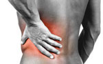
Back strain
Back strain is a fairly broad category called “soft tissue injury,” which covers muscles, tendons and ligaments. About 80% of back and neck pain is muscle-related.
The stomach muscles, or abdominals, enable the back to bend forward. They also assist in lifting. The abdominals work with the buttock muscles to support the spine. The oblique muscles go around the side of the body to provide additional support to the spine.
Another type of strain relates to spinal ligaments that run in front and in back of the vertebral bodies. Tendons, which also connect muscles in the spine, can develop inflammation, or tendonitis.
Some people believe that part of what makes the back muscles
more prone to strain is that they are shorter than other big muscles
in the body. The muscles in our thighs that enable us to walk, run and
jump are longer and less prone to strain. It’s very unusual to
strain a thigh muscle.
[top]
Fractures from Trauma
Spinal fractures are different than a broken arm or leg. A fracture of a spine vertebra can cause bone fragments to damage spinal nerves or the spinal cord.
A spinal fracture can occur from a fall, a car accident, or when an object impacts the spinal vertebrae with a force that it cannot withstand, so the bone cracks.
The most common type of spine fracture is a vertebral body compression fracture, which is downward force that shatters the structure of the vertebrae. If the force is great enough, it may send bone fragments into the spinal canal, called a burst fracture.
Most spinal fractures occur from severe trauma to the body from car accidents, falls, and other high impact force to the body. Injuries can range from spine vertebra fractures or as serious as debilitating spinal cord damage that can cause permanent paralysis.
Some smaller fractures can heal with physical therapy treatment and rest, however severe fractures will require surgery to reposition the bones and can include paralysis if the injury damaged the spinal cord. Spinal fractures and dislocations can pinch, compress, and even tear the spinal cord.
Fractures can occur anywhere along the spine, including the cervical, thoracic, and lumbar areas of the spine.
Spinal deformity
Skeletal irregularities place strain on the vertebrae and supporting muscles, tendons, ligaments and tissues supported by spinal column. These irregularities include scoliosis, a curving of the spine to the side; kyphosis, in which the normal curve of the upper back is severely rounded; lordosis, an abnormally accentuated arch in the lower back; back extension, a bending backward of the spine; and back flexion, in which the spine bends forward.
Spinal deformities can affect people of all ages but are most common in adolescents. The cause of these conditions are unknown, but abnormal bone and muscle growth are thought to be a contributing factor.
Spinal deformities become serious when they progress and threaten to cause severe pain and/or permanent disability. In other cases, some people with spinal deformity may not even know they have it. Treatment of these conditions aims at minimizing progression of the disease and preventing further growth.
Herniated Disc
The spine is composed of many vertebrae stacked on top of each other. Between these bones are discs, which act as shock absorbers. The shock-absorbing discs resemble jelly donuts, each having a jelly-like center. As we age, the discs naturally become less flexible and more brittle. Normal disc degeneration which naturally occurs with old age, can also cause pain.
Discs can herniate in any direction — forward, centrally or, most commonly, backward and sideways in the direction of the spinal nerves.
Herniated discs account for a small percentage of back pain.
While herniated discs are often referred to as “slipped discs,” this really isn’t accurate because discs don’t ever slip out of position. They are actually attached by connective tissue to vertebrae above and below. A disc herniation can be “contained” or “uncontained.” With a bulge, for example, the jelly center remains within the disc wall. "Uncontained" means the jelly center has broken through the annulus wall but stays connected to the nucleus pulposus. Or the herniation can be “sequestered,” when it breaks free from the nucleus and travels away from the disc.
Bulging Disc
While herniated discs are often referred to as “slipped discs,” this really isn’t accurate because discs don’t ever slip out of position. They are actually attached by connective tissue to vertebrae above and below. A disc herniation can be “contained” or “uncontained.” With a bulge, for example, the jelly center remains within the disc wall. "Uncontained" means the jelly center has broken through the annulus wall but stays connected to the nucleus pulposus. Or the herniation can be “sequestered,” when it breaks free from the nucleus and travels away from the disc.
A bulging disc forms when the wall of the disc is deformed but not necessarily herniated. The nucleus is still contained in the wall. You NEVER need surgery to treat a bulging disc.
Stenosis
Stenosis is a condition that can develop as a person ages, particularly in those over 50. It is characterized by a narrowing of the spinal canal, which places pressure on the spinal cord and nerves, because there is not enough room for them. It resembles placing a ring on your finger. If the finger becomes injured or inflamed, the ring constricts and causes pain. The pain caused by stenosis is typically focused in the low back area and can shoot down the legs and flare up after walking or exercising.
Spinal Tumor
Spinal cord tumors are abnormal growths of tissue found inside the bony spinal column, which is one of the primary components of the central nervous system (CNS). Benign tumors are noncancerous, and malignant tumors are cancerous. The CNS is housed within rigid, bony quarters (i.e., the skull and spinal column), so any abnormal growth, whether benign or malignant, can place pressure on sensitive tissues and impair function. Tumors that originate in the brain or spinal cord are called primary tumors.
Spondylolysis
Spondylolysis relates to instability of specific bones in the low back. It a very common cause of back pain, particularly in adolescents. Gymnasts who perform routines that bend and arch the back are often victims of spondylolysis or spondylolisthesis.
Spondylolisthesis and spondylolysis are caused by joint instability in the low back. The rear part of spinal vertebrae has facet joints that act as hinges, allowing our spines to twist and bend. Sometimes, however, the posterior element can crack. Either from heredity or wear and tear, part of the posterior element called the pars interarticularis can crack, causing the vertebrae to slip forward out of its correct position. Spondylolysis occurs when the PARS hinge is cracked, but the vertebrae is still in its correct position. Spondylolisthesis occurs when the cracked PARS has allowed the vertebrae to slide forward out of its correct position. If left untreated, spondylolysis can lead to spondylolisthesis.
Kphosis
Kyphosis and lordosis are types of spinal deformities. While slight curvature of the spine is normal and healthy, there are some cases where it is over-pronounced and can cause both cosmetic deformity and health risks. When the spine curves inward too much in the low back, it is called lordosis. When the spine in the shoulder blade or mid-spine area has too much forward curve, or too much of a hump, it is called kyphosis. Kyphosis most often occurs in the thoracic area of the spine.
Scoliosis
Scoliosis is typically a painless condition that causes an abnormal curvature in the spine. This will often create a rotation of the spine and rib cage, which creates asymmetry of the shoulders, trunk, and waist. Although most cases of scoliosis are mild, severe scoliosis can often be disabling. Certain conditions can help cause scoliosis such as cerebral palsy, but in most cases the cause of scoliosis is unknown.
The three main types of scoliosis are Functional, Neuromuscular, and Degenerative. Functional scoliosis is where the spine is normal, but eventually an abnormal curve starts to develop due to another aliment in the patient’s body such as having one leg longer than the other. Neuromuscular scoliosis can usually be much more severe since the condition is present at birth. This type of scoliosis is due to the failure of the spine bones to properly form, or fail to correctly separate from each other. People born with birth defects or cerebral palsy can often have neuromuscular scoliosis. Degenerative scoliosis is found in adults where the weakening of normal ligaments and other soft tissues of the spine can lead to an abnormal curve in the spine. Many times this can be linked to the patient having complained about arthritis. Idiopathic scoliosis is not linked to a known cause, but it is the most common type of scoliosis in adolescents. Doctors are unsure what causes this type of scoliosis, but it is suggested that it is hereditary because the disorder tends to run in families.
Degenerative Disc Disease
Degenerative disc disease commonly occurs with age, as discs become more brittle, less resilient and more prone to herniation. Degenerative disc disease is the single most common diagnosis related to serious back and neck pain. When a disc herniates in the spine, the surgeon can sometimes simply remove a portion of the disc. In other cases, where the disc is more damaged and must be removed, something must be placed into the disc space. Otherwise, the two vertebrae will collapse on top of one another, placing pressure on the nerve roots that branch off from the spinal cord.
Diagnosis
Outlined below are some of the diagnostic tools that your physician may use to gain insight into your condition and determine the best treatment plan for your condition.
- Medical history: Conducting a detailed medical history helps the doctor better understand the possible causes of your back and neck pain which can help outline the most appropriate treatment.
- Physical exam: During the physical exam, your physician will try to pinpoint the source of pain. Simple tests for flexibility and muscle strength may also be conducted.
- X-rays are usually the first step in diagnostic testing methods. X-rays show bones and the space between bones. They are of limited value, however, since they do not show muscles and ligaments.
- MRI (magnetic resonance imaging) uses a magnetic field and radio waves to generate highly detailed pictures of the inside of your body. Since X-rays only show bones, MRIs are needed to visualize soft tissues like discs in the spine. This type of imaging is very safe and usually pain-free.
- CT scan/myelogram: A CT scan is similar to an MRI in that it provides diagnostic information about the internal structures of the spine. A myelogram is used to diagnose a bulging disc, tumor, or changes in the bones surrounding the spinal cord or nerves. A local anesthetic is injected into the low back to numb the area. A lumbar puncture (spinal tap) is then performed. A dye is injected into the spinal canal to reveal where problems lie.
- Electrodiagnostics: Electrical testing of the nerves and spinal cord may be performed as part of a diagnostic workup. These tests, called electromyography (EMG) or somato sensory evoked potentials (SSEP), assist your doctor in understanding how your nerves or spinal cord are affected by your condition.
- Bone scan: Bone imaging is used to detect infection, malignancy, fractures and arthritis in any part of the skeleton. Bone scans are also used for finding lesions for biopsy or excision.
- Discography is used to determine the internal structure of a disc. It is performed by using a local anesthetic and injecting a dye into the disc under X-ray guidance. An X-ray and CT scan are performed to view the disc composition to determine if its structure is normal or abnormal. In addition to the disc appearance, your doctor will note any pain associated with this injection. The benefit of a discogram is that it enables the physician to confirm the disc level that is causing your pain. This ensures that surgery will be more successful and reduces the risk of operating on the wrong disc.
- Injections: Pain-relieving injections can relieve back pain and give the physician important information about your problem, as well as provide a bridge therapy.